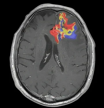
Our automated software leverages exclusive quantitative technology to accurately characterize brain tumors
FTB Maps
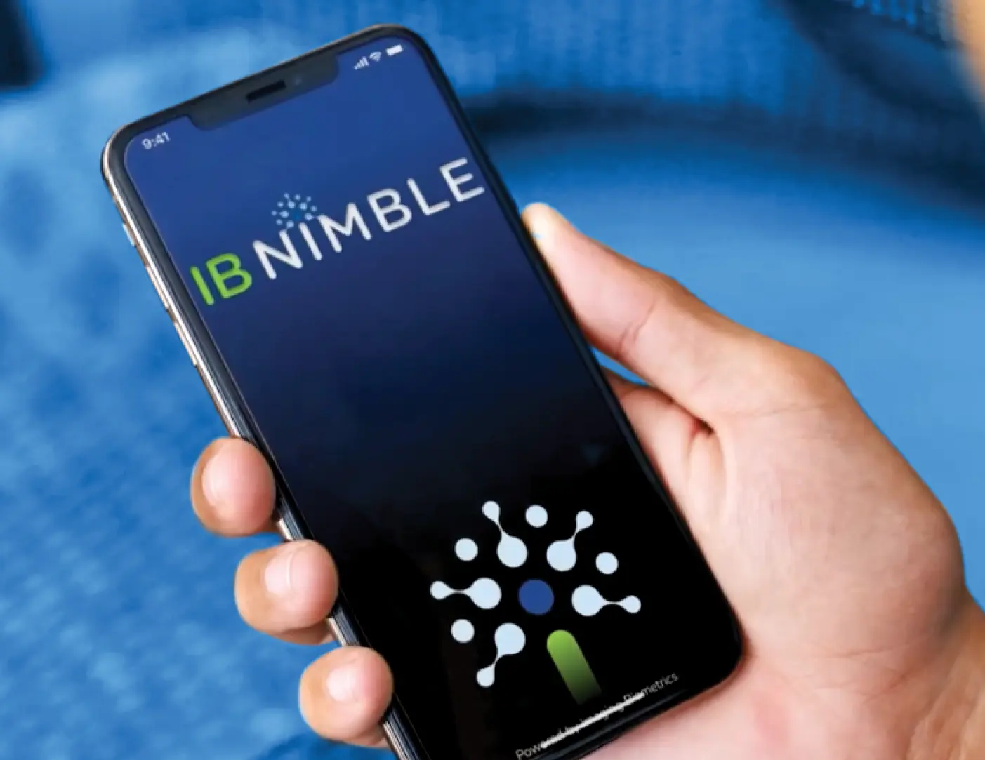
IB Nimble
Expanded Access Program

When our clients come to us in search of help with difficult problems, they know that they can count on us to provide new insights and meaningful improvements, always with patient care as our highest priority. Through our close collaborations with top research minds in the medical imaging field, we help translate proprietary and sophisticated advancements into easy-to-use, intuitive software solutions for clinicians and researchers. Our mission is to help you get the most meaning out of your medical imaging studies. Our commitment is to serve your patients by bringing only proven solutions to market, giving you assurance that when you are relying on our solutions, your team is providing the best care possible.
In The News
By providing accurate and intuitive information to clinicians, optimal treatment decisions are made faster and with greater confidence.
proven